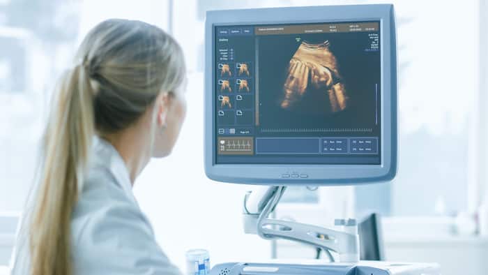
Ultrasound Imaging at IMPC Paris
Ultrasound in Paris
Seeking for an Ultrasound exam in Paris
Introduction to Ultrasound Imaging: A Safe and Non-Invasive Diagnostic Tool
Ultrasound imaging, also known as sonography, leverages high-frequency sound waves to generate visual representations of the interior of the body. This advanced technology stands out for its ability to capture real-time images, allowing not only the observation of internal organs but also the movement of blood through vessels, without exposing patients to ionizing radiation—a significant advantage over traditional X-ray imaging.

How Ultrasound scan Works: The Science Behind the Images
At the core of ultrasound exams lies the transducer, or probe, which is applied directly onto the skin or inserted into a body opening. A special gel applied on the skin ensures the ultrasound waves travel efficiently from the transducer through the gel and into the body. The images are then created based on the reflection of these waves off bodily structures. The amplitude of the sound signal and the time it takes for the waves to travel through the body are critical in producing a detailed image.
Diverse Applications of Ultrasound scan
This exam serves as a pivotal medical tool across various specialties. Its applications range from abdominal and breast imaging to more specialized uses such as bone sonometry, Doppler fetal heart rate monitoring, and even guiding biopsies. This versatility underscores ultrasound’s role in diagnosing and treating a wide array of medical conditions.
Safety Profile and Prudent Use
With over two decades of use, ultrasound imaging boasts an exemplary safety record, attributed to its reliance on non-ionizing radiation. However, like any medical procedure, it necessitates prudent use by trained health professionals to minimize any potential biological effects on the body, such as slight tissue heating or cavitation. Special caution is advised during pregnancy, with recommendations against non-medical uses like keepsake videos, to avoid unnecessary exposure.
Innovations in Ultrasound: From 3D to 4D Imaging
The evolution from two-dimensional (2D) to three-dimensional (3D) and even four-dimensional (4D) ultrasound has revolutionized prenatal care, offering detailed visualizations of the fetus. Despite the safe profile of ultrasound, the FDA cautions against non-medical 3D ultrasounds for keepsake purposes due to potential risks from prolonged exposure.
Who Performs Exams?
Ultrasound examinations are conducted by doctors or specialized healthcare professionals known as sonographers or ultrasound technicians. Their expertise ensures the safe and effective operation of ultrasound machines, emphasizing the importance of seeking care from qualified medical personnel in appropriate settings.
Ultrasound Imaging Appointment
Take an appointment for untrasound exam in our centers:
IRM Bachaumont 75002
Preparing for Your Ultrasound: What to Expect
Preparation for an ultrasound varies depending on the type. Some require no prior preparation, while others may necessitate dietary adjustments or a full bladder.
At the outset of an ultrasound examination, a crucial step involves the application of a specialized gel to the skin surface covering the targeted area. This gel plays a pivotal role in the diagnostic process, serving as a medium that eliminates air gaps which could otherwise impede the transmission of ultrasound waves. Formulated from a safe, water-based composition, this gel is designed for easy application and removal, ensuring it does not leave residues on the skin or clothing.
The Role of the Sonographer and the Transducer
Central to the ultrasound procedure is the expertise of a trained technician, known as a sonographer, who employs a hand-held device called a transducer. This critical instrument is meticulously pressed against the specified area and maneuvered as necessary to procure optimal images. It operates by emitting sound waves into the body, capturing the echoes that rebound, and conveying these signals to a computer. This sophisticated system then interprets these echoes into images that offer a glimpse into the body’s interior landscape.
Innovations in Internal Ultrasound Techniques
Ultrasound technology has evolved to include procedures that allow for internal examination, providing even deeper insights into the body’s internal health. These specialized techniques involve attaching the transducer to a probe, which is then carefully inserted into a natural orifice to target specific internal structures. Among these advanced methods are:
- Transesophageal Echocardiogram: This procedure, conducted under sedation, involves the insertion of a transducer through the esophagus to acquire detailed images of the heart.
- Transrectal Ultrasound: Aimed at evaluating the prostate, this technique employs a specially designed transducer introduced into the rectum.
- Transvaginal Ultrasound: For a closer examination of the uterus and ovaries, a specialized transducer is gently inserted into the vagina.
Duration of the Procedure
A typical ultrasound session is characterized by its efficiency and brevity, usually completed within a timeframe ranging from 30 minutes to an hour. This expedient nature does not detract from the procedure’s ability to provide comprehensive and detailed insights, making it a highly valued tool in medical diagnostics.
Understanding Your Results
The timeframe for receiving ultrasound results can vary. In some instances, such as prenatal ultrasounds, immediate analysis and feedback are possible. Otherwise, a radiologist will review the images and relay findings to your healthcare provider. This exam can detect a wide range of conditions, from abnormal growths and blood clots to pregnancy-related issues and organ-specific ailments.
Why choosing IMPC for an Ultrasound Exam ?
IMPC (Imagerie Médicale Paris Centre) offers a comprehensive range of diagnostic imaging services in the heart of Paris. Here are some compelling reasons to consider IMPC for your ultrasound exam:
- Advanced Technology: IMPC boasts a state-of-the-art facility equipped with the latest technologies in conventional radiology, interventional radiology, 3D EOS imaging, ultrasound, MRI, CT scans, and bone densitometry. Their cutting-edge equipment ensures accurate and detailed imaging.
- Expert Specialists: IMPC houses a team of specialized medical professionals who excel in various diagnostic fields. Whether you need an abdominal, pelvic, obstetrical, pediatric, hip or joint ultrasound, their experts provide precise interpretations.
- Integrated Services: With an on-site integrated laboratory, IMPC’s radiologists can review your biological analysis results in real-time during your clinical examination. This seamless integration allows for comprehensive assessment and personalized recommendations.
- Flexible Appointments: IMPC’s imaging centers are open from Monday to Friday, 8:00 AM to 8:00 PM. You can conveniently schedule appointments based on your availability.
- Wide Range of Exams: IMPC offers a variety of diagnostic exams, including:
- MRI (Magnetic Resonance Imaging): Detailed imaging without X-rays, using magnetic fields.
- CT Scan (Computed Tomography): 2D or 3D X-ray images for precise anatomical visualization.
- Ultrasound: Non-invasive imaging using sound waves.
- Mammography: Specialized X-rays for breast imaging.
- Financial Assistance: IMPC understands that healthcare costs can be a concern. They offer financial assistance options for patients without insurance coverage.
Whether you’re seeking routine screenings or investigating specific health concerns, IMPC’s expertise and commitment to patient care make it a reliable choice for ultrasound exams in Paris.
