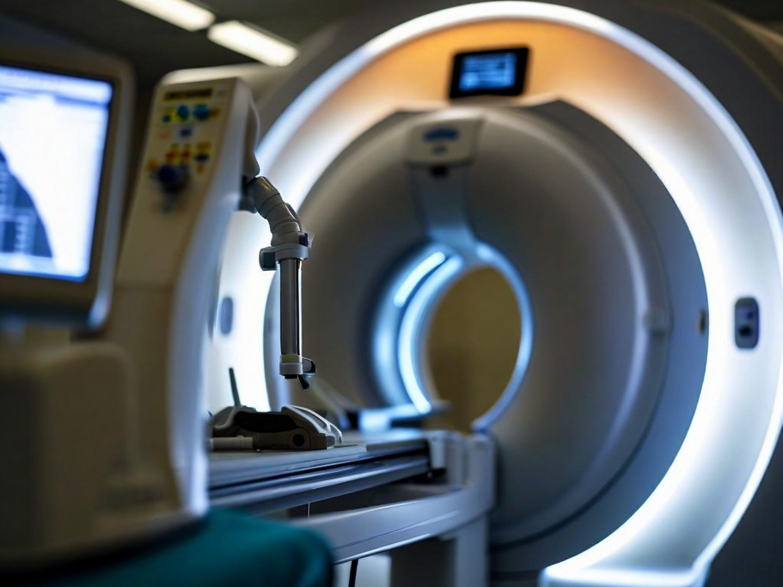
Heart ct scan or cardiac ct scan
Heart ct scan in Paris
RDV scanner en ligne sur Doctolib
Prenez rendez-vous pour une IRM dans nos centres
Rendez-vous Doctolib IMPC Bachaumont – IRM Paris 75002
Rendez-vous Doctolib Pôle Santé Bergère – IRM Paris 75009
Rendez-vous Doctolib Clinique Blomet- IRM Paris 75015

The heart ct scan also known as cardiac ct scan play a crucial role in the meticulous examination of the human heart. The body is an indispensable organ that requires vigilant observation for lasting well-being. In this exploration of cardiovascular imaging we shed light not only on the usefulness of a heart ct scan and the subtleties of different cardiac scanning modalities. From traditional X-ray and CT images to advanced techniques such as molecular imaging and nuclear medicine, the range of imaging services available in a modern medical center is expanding. This article looks at the various methodologies employed in cardiovascular imaging, highlighting the subtleties of emission-based technologies and their essential role in comprehensive assessment of the heart.
Coronary CT scanner: what is it used for?
A coroscanner enables detailed examination of the coronary arteries and heart chambers. It plays a key role in the early diagnosis of coronary artery disease, detecting blockages, narrowings and malformations of the blood vessels. This procedure is often used to assess cardiac risk, monitor the evolution of existing pathologies and aid medical decision-making.
Thanks to its ability to provide detailed cross-sectional images of the heart, the coronary scanner offers precise analysis of cardiac structures, enabling healthcare professionals to effectively diagnose a variety of conditions, including coronary artery disease.
Why have a cardiac ct scan ?
A decision to have a heart ct scan stems from a proactive approach to monitoring and maintaining optimum heart health. From a medical standpoint, people choose this test for its diagnostic prowess in assessing various aspects of cardiac well-being. These examinations, often performed on an outpatient basis, involve qualified technologists using advanced imaging techniques, such as cross-sectional imaging and injection of contrast agents if necessary.
A cardiac ct scan provides a complete examination of the heart's conditionThis enables healthcare professionals, particularly cardiologists, to accurately diagnose and manage potential problems. Thanks to cross-section and à detailed soft-tissue imagingthese scanners provide invaluable information about the structure and function of the heart. Whether they are used to assess known risk factors, to investigate symptoms or as part of routine examinations, the heart ct scan offer a comprehensive, non-invasive means of assessing heart health, contributing to early diagnosis and effective medical intervention where necessary.
Coronary CT scan with injection: a precise examination
A coronary scanner is an advanced technique for obtaining detailed images of the coronary arteries and heart muscle. This iodinated contrast medium, injected intravenously, enhances the visibility of cardiac structures and helps detect abnormalities such as obstructions or narrowing of the coronary arteries. This rapid, non-invasive examination is particularly effective in diagnosing coronary artery disease and guiding therapeutic decisions.
What are the steps involved in cardiac imaging?
The imaging produced by the coronary scanner provides a detailed map of your coronary arterieshighlighting blockagesthe plates or the anomalies. It goes beyond the surface, delving into the very fabric of your heart's health. This comprehensive overview enables healthcare professionals to make informed decisions about your cardiovascular well-being.
Coronary Scanner imaging calls on cutting-edge technologies such as computed tomography (CT) or imaging by magnetic resonance (MRI). These methods allow detailed visualization of the heart's anatomy, enabling health professionals to identify any abnormalities.
Un outil radiologique de pointe pour l’imagerie diagnostique cardiaque
Le coroscanner, ou scanner cardiaque, est un examen radiologique de haute précision qui permet de visualiser les artères coronaires avec une finesse remarquable. Réalisé au sein d’un service d’imagerie ou d’un centre hospitalier, il repose sur la tomographie assistée par ordinateur et, selon les indications, peut nécessiter une injection de produit iodé. Encadré par un médecin radiologue et un manipulateur, l’examen est rapide, indolore, et expose le patient à une faible irradiation, strictement contrôlée. Utilisé en imagerie diagnostique, ce scanner permet la détection précoce de pathologies cardiovasculaires sans recours à une ponction ou à une anesthésie. En complément des radiographies conventionnelles, il offre une exploration complète du thorax et du corps humain, et s’avère indispensable pour guider certaines prises en charge ou anticiper des complications. Grâce à ses détecteurs modernes et à l’usage raisonné des produits de contraste, le coroscanner s’impose comme une référence incontournable parmi les examens d’imagerie cardiaque.
How does a cardiac scan work?
The cardiac ct scan procedure itself is completely painless and non-invasive.. The patient lies on a comfortable, specialized table while the the scanner rotates around his chest and captures detailed images. This approach resembles a meticulous scanning session focused on diagnosing internal organs, eliminating the need for product injections or discomfort.
The process is fast and involves minimal exposure to radiation. The medical team skilfully guides patients through each step, prioritizing both comfort and precision. The emphasis on diagnosing internal organs, particularly those related to the vascular health, underscores heart ct scans ability to provide precise cardiovascular assessments.
What are the risks of this examination?
A coroscanner is a low-risk procedure, but some people may experience problems with the contrast medium or other substances used during the examination.
Contrast medium
Some people have an adverse reaction to the contrast medium, which usually contains iodine. After injection, symptoms such as itching, nausea, sneezing or a rash may occur. These symptoms usually disappear without treatment, but antihistamines, steroids and histamine blockers may be helpful.
If you have diabetes or kidney disease, you may need extra fluids after your examination. This will help eliminate iodine from your body.
Since the contrast material used in a cardiac scan can pass into your breast milk, it may be a good idea to prepare some milk before your examination to give to your baby for a day or two afterwards.
Write-off
Scanners use X-rays. Exposure to this radiation carries a low long-term risk of cancer. For your safety, the amount of radiation exposure is minimized. However, since X-rays can harm a developing fetus, this procedure is not recommended if you are pregnant. If you are scheduled for a cardiac CT scan, your healthcare professional can take steps to protect your baby.
Qu’est-ce qu’un coroscanner ?
A coroscanner is a medical imaging technique that uses X-rays to produce high-resolution images of the heart and coronary vessels. Sometimes, a contrast agent is injected to improve visualization of arteries and heart tissue, enabling a more complete assessment. The use of iodinated contrast enhances visualization of suspicious areas, facilitating diagnosis.
Dernière mise à jour : le 30 mai 2025
Controlled by Dr Antoine Hakime
