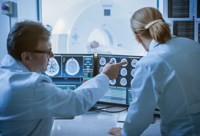
How does an MRI scan work?
Découvrez comment se passe une IRM à chaque étape de l’examen.
What is an MRI scan?
MRI, or Magnetic Resonance Imaging, is a technique that enables radiologists to see into the human body in great detail without using X-rays. MRI images are obtained using a powerful magnetic field, radio waves and a complex computer system. The technique is safe and painless.
Le déroulement de l’examen: comment se passe une IRM ?
À votre arrivée, on vous posera, comme lors de la prise du rendez-vous, plusieurs questions ; le plus important est de signaler que vous n’avez ni pile cardiaque (pacemaker), ni valve cardiaque, ni d’élément contenant du fer près des yeux ou dans la tête. Cela est crucial, car l’IRM est un examen utilisant un champ magnétique puissant, et toute présence de métal peut interférer avec le processus.
Pour permettre d’obtenir des images de bonne qualité, on vous indiquera les vêtements que vous devrez enlever. L’IRM se comporte comme un aimant géant vis-à-vis des objets contenant certains métaux. Vous ne garderez donc aucun bouton, agrafe, barrette de cheveux ou fermeture éclair métallique. Vous laisserez au vestiaire, dans un casier, vos bijoux, montre, clefs, porte-monnaie, cartes à bande magnétique (carte de crédit, de transport, etc.) et votre téléphone portable.
Vous entrerez dans une salle qui sera fermée pendant l’examen d’imagerie.
How does an MRI scan work?
Vous serez allongé sur une table d’examen qui se déplace dans une sorte de tunnel pour la plupart des appareils, généralement sur le dos et seul dans la salle d’examen. Nous communiquerons avec vous grâce à un micro. Dans tous les cas, l’équipe se trouve tout près de vous, derrière une vitre. Elle vous voit et vous entend pendant tout l’examen.
Si vous voulez nous appeler, vous pourrez utiliser une sonnette que l’on placera dans votre main. Si cela est nécessaire, on peut à tout moment intervenir ou interrompre l’examen. Vous resterez en moyenne 10 à 20 minutes dans la salle d’examen. Votre coopération est essentielle : vous devez essayer de rester parfaitement immobile ; dans certains cas, nous vous dirons, à l’aide du micro, quand arrêter de respirer pour quelques secondes.
À cet instant précis, vous entendrez un bruit répétitif, comme celui d’un moteur de bateau ou d’un tam-tam, pendant ce qu’on appelle une séquence. Un examen contient généralement de 3 à 6 séquences, voire plus si nécessaire. L’IRM est un examen particulièrement utile pour visualiser des tissus mous, comme les muscles, les ligaments ou les organes internes. Dans certains cas, des examen d’imagerie supplémentaires peuvent être réalisés pour affiner le diagnostic, notamment pour les pathologies ostéo-articulaires. Pour diminuer le bruit et rendre l’examen plus agréable, vous pourrez parfois utiliser des bouchons d’oreille ou même écouter de la musique pendant l’examen.
Some tests require an intravenous injection, usually at the elbow.

Is MRI dangerous to health?
Que ressentirez-vous pendant une IRM ?
The examination is not painful, but it is often a little long and the noise can be unpleasant.
A feeling of discomfort due to fear of being locked in (claustrophobia) is a common problem that
we know well. It can often be reduced by simple means, without any treatment.
If, for example, you feel uncomfortable in an elevator, tell the reception staff right away so that they can take special care of you.
Why does an MRI take so long?
An MRI scan consists of several sequences (4 to 5 in general), each lasting 3-4 minutes on average, to ensure good image quality; this enables organs to be viewed in several spatial planes (axial, sagittal, frontal, etc.), with different tissue contrasts (T1- or T2-weighted images). Some sequences are repeated after contrast medium injection. Image acquisition therefore takes an average of 20 minutes, to which must be added the patient's welcome, explanations to the patient, undressing, time to install the machine, time to inject the product, uninstalling the patient, and pauses between sequences. If the patient moves during a sequence, it will have to be repeated, which will lengthen the examination.
How long does an MRI examination take?
The average MRI scan lasts between 15 and 20 minutes. Some may be shorter, others longer.
What does an MRI scan look like?

Résultats IRM: que faire ensuite ?
An injection for an MRI: how and what are the risks?
The most commonly used contrast medium is Gadolinium.
This product is generally well tolerated. Trivial allergic reactions (urticaria) are possible.
Very serious allergic reactions are quite exceptional.
The sting may cause a small hematoma, which is not serious and will spontaneously heal within a few days.
During injection, pressure may cause the product to leak under the skin, into the vein.
This complication is rare (one case in several hundred injections, generally without serious consequences), and may exceptionally require local treatment.
Why is an MRI scan expensive?
The device itself requires a large investment for its acquisition and installation (electromagnetic shielding), and incurs very high annual maintenance costs. The lifetime of a device is around 7 years. Highly qualified personnel are required, as well as frequent updating of the entire computer system and software. An MRI technician performs your examination, and therefore spends between twenty minutes and half an hour per patient. Finally, image interpretation requires in-depth, specialized training on the part of the radiologist, as the technique is quite complex. The high cost of MRI is offset by the very high performance of this technique, enabling you to avoid other more complex or painful examinations to reach a diagnosis.
IRM: De quoi s’agit-il ?
MRI stands for Magnetic Resonance Imaging.
Le mot magnétique indique que l’appareil comporte un gros aimant ; le mot résonance indique que l’on va utiliser des ondes de radiofréquence, comme celles des téléphones portables pour faire vibrer les nombreux noyaux d’hydrogène composant les tissus de votre corps, et fabriquer ainsi des images. L’IRM est une procédure sécurisée et non invasive, essentielle pour poser des diagnostics précis. Si vous vous demandez comment se passe une IRM ou avez des questions concernant vos résultats IRM, n’hésitez pas à consulter un de nos professionnels de santé. Chaque étape de l’examen est conçue pour garantir votre confort et la qualité des images.
Noise during sequences
Noise is caused by the rapidly alternating current flowing through the coils that create the radio waves required for image acquisition.
Je suis allergique: que faire lors d'une IRM?
Please bring with you on the day of the exam
After your return home
In the vast majority of cases, you won't feel anything in particular. However, don't hesitate to let the team know if anything seems out of the ordinary.
It's normal to have questions about the exam you're about to take.
We hope we've answered your questions. Please do not hesitate to contact us again should you require any further information.
MRI and pacemakers?
Patients with pace-makers are not allowed inside the MRI chamber, as the magnetic field will disrupt the battery, causing heart rhythm disturbances that can be severe.


