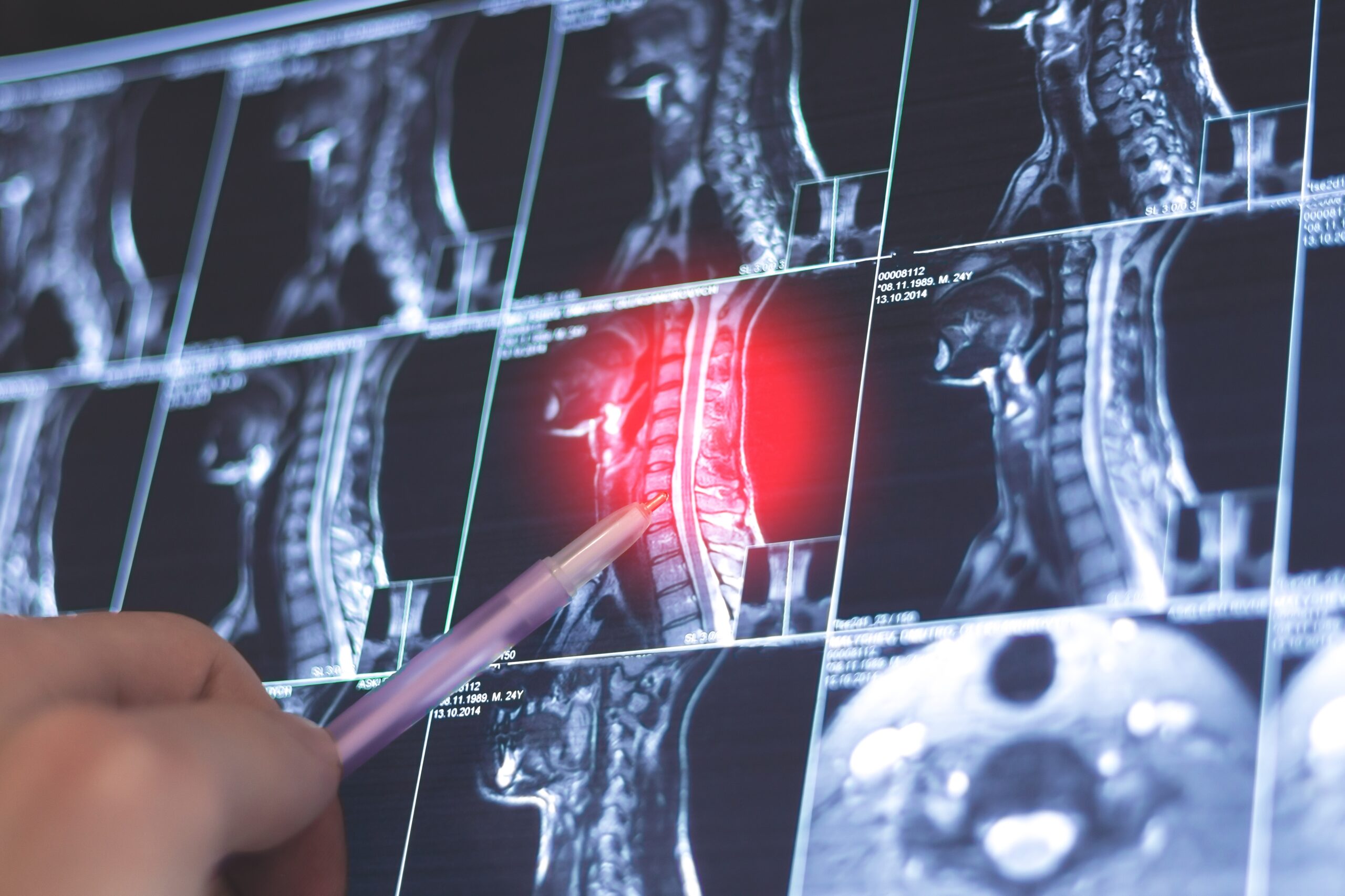
MRI of the brachial plexus
Brachial plexus MRI in the neurology department
RDV IRM en ligne sur Doctolib
Prenez rendez-vous pour une IRM dans nos centres
Rendez-vous Doctolib IMPC Bachaumont – IRM Paris 75002
Rendez-vous Doctolib Clinique Drouot – IRM Paris 75009
Rendez-vous Doctolib Pôle Santé Bergère – IRM Paris 75009
Rendez-vous Doctolib Clinique Blomet- IRM Paris 75015

L’IRM du plexus brachial is a advanced medical imaging technology used to evaluate the nerve structures of the brachial plexus, an essential anatomical region of the peripheral nervous system. This diagnostic procedure provides detailed images and valuable information on pathologies that can affect the brachial plexusThis enables doctors to make accurate diagnoses and develop appropriate treatment plans. In this article, we take a closer look at theIRM du plexus brachial et expliquer les différentes pathologies qui peuvent être identifiées grâce à ce diagnostic.
Qu’est-ce que l’IRM du plexus brachial ?
L’IRM (imagerie par résonance magnétique) du plexus brachial est une procédure d’imagerie non invasive qui utilise des champs magnétiques et des ondes radio pour créer des images détaillées du plexus brachial. Le plexus brachial est un réseau de nerfs situé dans la région du cou et de l’épaule qui contrôle les mouvements et la sensibilité des membres supérieurs, y compris les bras, les épaules et les mains. L’IRM du plexus brachial permet d’obtenir des informations précises sur l’anatomie du plexus brachial et d’identifier les éventuelles anomalies ou pathologies qui pourraient affecter son fonctionnement.
Pourquoi l’IRM du plexus brachial est-elle utilisée ?
L’IRM du plexus brachial est utilisée pour évaluer diverses pathologies et affections touchant cette région du corps. Les principales raisons d’effectuer une IRM du plexus brachial sont :
-
Trauma assessment : L’IRM du plexus brachial can help detect nerve damage, nerve breaks or tissue tears that may occur as a result of trauma, such as a fall or car accident.
-
Tumor diagnosis : L’IRM du plexus brachial allows us to visualize tumors growing in the brachial plexus and assess their size, location and extent.
-
Évaluation des neuropathies : L’IRM du plexus brachial can help identify peripheral neuropathies, such as diabetic neuropathy, compressive neuropathy or inflammatory neuropathy, which can cause pain, numbness or weakness in the upper limbs.
-
Diagnosis of thoracic outlet syndrome : L’IRM du plexus brachial is used to diagnose thoracobrachial outlet syndromes, a condition in which the blood vessels or nerves of the brachial plexus are compressed as they pass between the clavicle and the first rib.
-
Assessment of demyelinating diseases : L’IRM du plexus brachial can help detect demyelinating diseases, such as multiple sclerosis, which can affect the brachial plexus and cause neurological symptoms.
Atteintes neurologiques et compléments d’exploration du plexus brachial
L’IRM du plexus brachial permet de détecter des pathologies du système nerveux périphérique et d’orienter la prise en charge des patients souffrant de troubles moteurs, sensitifs ou inflammatoires. Elle identifie les atteintes des axones, des racines rachidiennes ou du tronc nerveux, et peut être couplée à un électromyogramme (EMG) pour évaluer la contraction musculaire, les réflexes et l’état fonctionnel des nerfs moteurs. Certaines paralysies aiguës ou sensorielles peuvent résulter de lésions médullaires, vasculaires ou cervicales, provoquant une gêne dans les membres supérieurs, voire parfois dans les membres inférieurs. L’IRM aide ainsi à détecter des anomalies antérieures ou postérieures du canal rachidien, à analyser les effets secondaires liés à des maladies du système nerveux cérébral et à prévenir les suites neurologiques ou les séquelles durables en facilitant une stimulation et une rééducation ciblées.
Principales pathologies détectées par l’IRM du plexus brachial
L’IRM du plexus brachial est un outil précieux pour détecter différentes pathologies qui peuvent affecter cette région nerveuse essentielle. Voici quelques-unes des principales affections qui peuvent être identifiées grâce à l’IRM :
1. Tumeurs du plexus brachial
MRI can be used to visualize tumors that develop in the brain. brachial plexus, telles que les schwannomes ou les neurofibromes. Ces tumeurs peuvent entraîner des symptômes tels que des douleurs, des engourdissements ou des faiblesses dans les membres supérieurs. L’IRM fournit des informations précieuses sur la taille, l’emplacement et l’extension de la tumeur, ce qui est essentiel pour établir un plan de traitement adapté. Les tumeurs du plexus brachial peuvent se développer pour diverses raisons. Voici quelques-unes des principales causes associées à ces tumeurs :
-
Schwannomas : les schwannomes sont des benign tumours that form from Schwann cellswhich surround and insulate the peripheral nerves. These tumors can develop in the brachial plexus and cause symptoms such as pain, numbness or weakness in the upper limbs. Schwannomas can be sporadic, i.e. without a specific identifiable cause, or associated with hereditary conditions such as neurofibromatosis type 2.
-
Neurofibromas : les neurofibromes sont des tumors that form from nerve support cellscalled Schwann cells and fibroblasts. They can also develop in the brachial plexus, causing symptoms similar to those of schwannomas. Neurofibromas can be associated with neurofibromatosis type 1, a genetic disease characterized by the development of multiple nerve tumors throughout the body.
-
Metastatic tumors : dans certains cas, cancerous tumors from other parts of the body can spread (metastasis) and reach the brachial plexus. The cancers most commonly associated with brachial plexus metastases are lung cancer, breast cancer and kidney cancer. The identification of metastases in the brachial plexus may indicate an advanced stage of the disease, and requires further evaluation to determine the primary origin of the cancer.
-
Nerve trauma : bien que moins fréquentes, les tumours can develop following nerve trauma to the brachial plexus. Traumatic nerve damage can disrupt the normal growth and regeneration of nerve cells, leading to tumor formation.
2. Traumatic nerve damage
These trauma, such as car accidents or falls, can lead to nerve damage to the brachial plexus. MRI can visualize these lesions, determine their extent and provide detailed information on the nerve structures affected. This helps doctors to assess the severity of the lesion and draw up a plan for rehabilitation or surgical treatment if necessary.
3. Compressive neuropathy
A compressive neuropathy is a condition in which the nerves of the brachial plexus are compressed or trapped. The result is symptoms such as pain, numbness or weakness in the upper limbs. MRI can identify areas of compression and help doctors determine the underlying cause of compressive neuropathy, such as a tumor, herniated disc or enlarged muscle.
4. Thoracic outlet syndrome
These thoracobrachial outlet syndromes are characterized by compression of blood vessels or brachial plexus nerves between the clavicle and the first rib. MRI can help visualize this compression and identify the underlying cause, whether an anatomical anomaly, tumor or inflammation. This information is essential in determining the appropriate treatment plan, whether physiotherapy, medication or surgery.
5. Demyelinating diseases
Les maladies démyélinisantes du plexus brachial are conditions in which the myelin sheath surrounding nerve fibers in the brachial plexus is damaged. This can lead to impaired transmission of nerve signals and provoke various upper limb symptoms. Here are some of the main causes of demyelinating diseases :
- Multiple sclerosis : la sclérose en plaques est une maladie auto-immune dans laquelle le système immunitaire attaque la gaine de myéline du système nerveux central, y compris le plexus brachial. Les raisons exactes de cette réaction auto-immune ne sont pas encore entièrement comprises, mais des facteurs génétiques et environnementaux peuvent contribuer au développement de la maladie.
- Chronic inflammatory demyelinating neuropathy (CIDP) : la CIDP est une maladie démyélinisante inflammatoire chronique qui affecte les nerfs périphériques, y compris le plexus brachial. Les mécanismes exacts à l’origine de la CIDP ne sont pas encore totalement élucidés, mais il est suggéré qu’une réponse auto-immune anormale puisse jouer un rôle dans le développement de cette affection.
- Multifocal motor neuropathy (MMN) : la NMM est une maladie démyélinisante du système nerveux périphérique qui affecte principalement les nerfs moteurs. Bien que les causes exactes de la NMM ne soient pas encore complètement comprises, il est supposé que des anomalies du système immunitaire peuvent jouer un rôle dans son développement.
- Viral infections : certaines infections virales, telles que le virus d’Epstein-Barr, le cytomégalovirus et le virus de l’hépatite C, ont été associées à des cas de maladies démyélinisantes du plexus brachial. Il est suggéré que ces infections virales peuvent déclencher une réponse auto-immune qui endommage la gaine de myéline des nerfs du plexus brachial.
- Environmental factors : des facteurs environnementaux tels que l’exposition à certaines toxines, aux métaux lourds ou à des produits chimiques peuvent également jouer un rôle dans le développement des maladies démyélinisantes du plexus brachial. Cependant, la relation exacte entre ces facteurs environnementaux et les maladies démyélinisantes nécessite encore des recherches approfondies.
MRI can help detect signs of demyelination and assess the extent of lesions in the brachial plexus. This enables doctors to monitor disease progression and make informed decisions about treatment.
Conclusion
MRI of the brachial plexus is a key examination in the evaluation of neurological disorders affecting the nerves of the shoulder and arm. It allows us to analyze the spinal cord and detect any pathologies that may cause various symptoms, such as acute pain, muscle weakness or sensory disorders. Indeed, the nerves of the brachial plexus are responsible for motricity and sensation in the upper limbs, and any damage at this level can have major functional consequences for sufferers.
During the examination, a neurologist can identify signs of inflammation or neuronal degeneration, which may indicate pathologies such as Charcot's amyotrophic lateral sclerosis or traumatic lesions. MRI can also assess nerve conduction through the plexus trunk, providing valuable information on the state of the cranial nerves and their impact on motor skills and limb sensitivity. In cases of paralysis, this examination is essential to guide the choice of appropriate therapeutic treatments.
The MRI results also guide the rehabilitation steps that are essential to improve muscle function and quality of life for affected patients. A multidisciplinary approach, such as that implemented at the IMPC, involving neurologists, physiotherapists and other healthcare professionals, is often necessary to optimize the management of neurological disorders associated with brachial plexus damage. MRI thus represents a fundamental tool in the diagnosis and management of neuromuscular pathologies, facilitating the path to functional recovery.
Dernière mise à jour : le 30 mai 2025
Dr Françoise Heran
