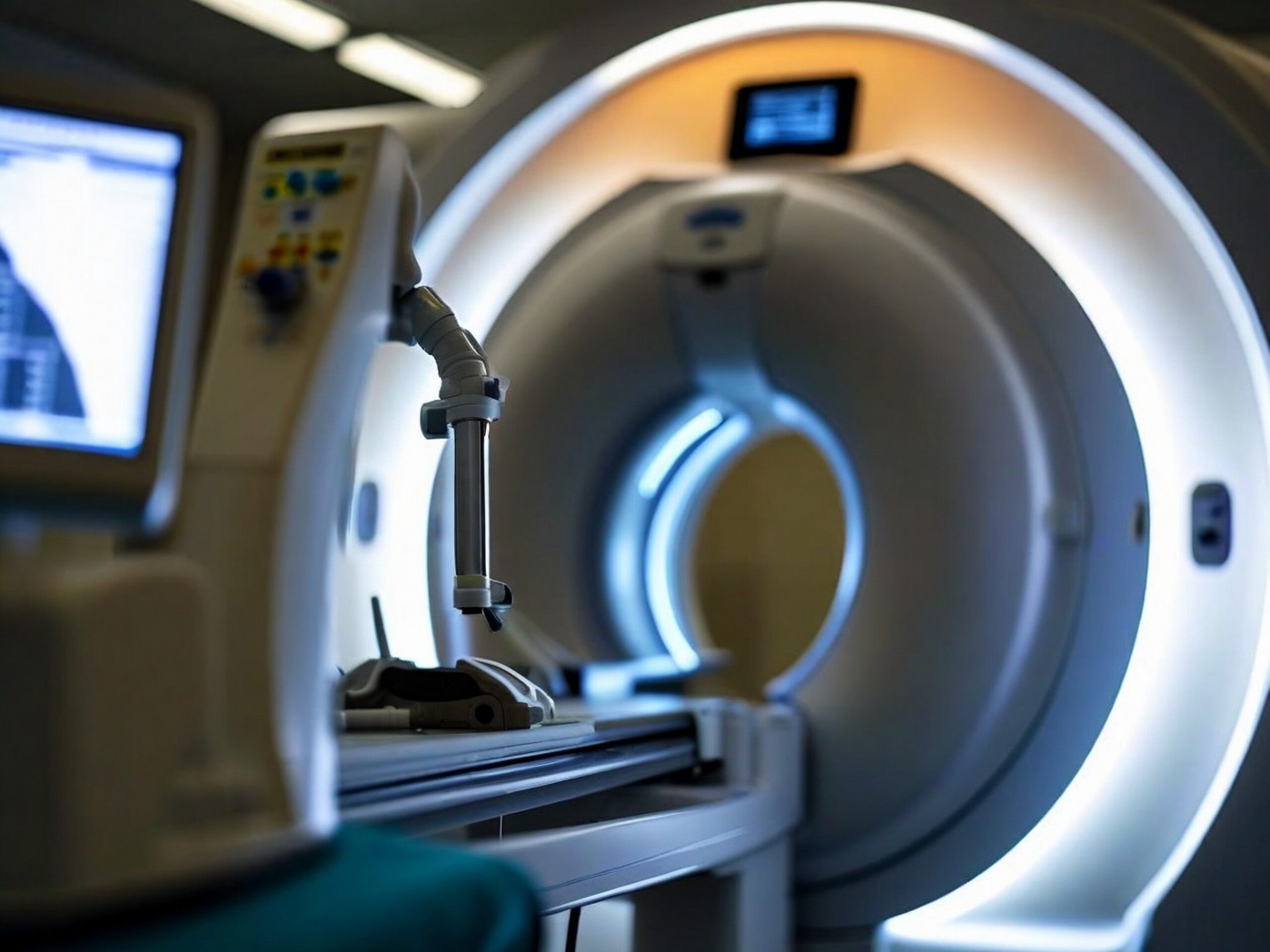
Arthroscanner : comment se déroule l’examen ?

RDV scanner en ligne sur Doctolib
Prenez rendez-vous pour un scanner dans nos centres
Prise de rendez-vous SCANNER – Paris 75002
Prise de rendez-vous SCANNER – Paris 75009
Prise de rendez-vous SCANNER – Paris 75015

Comment se passe un arthroscanner dans notre centre ?
L’examen se déroule en deux étapes. Le patient est allongé confortablement, et l’articulation est soigneusement désinfectée avant l’injection.
La particularité de nos centres IMPC est de proposer un accompagnement personnalisé : le manipulateur-radio et le médecin radiologue vous guident à chaque étape de la procédure, afin de garantir confort et sécurité.
Quelles sont les étapes de l’examen ?
Accueil et préparation : installation en cabine, retrait de tout objet métallique, explication du déroulé.
Injection : sous contrôle radiographique, le radiologue injecte un produit iodé dans l’articulation. Dans certains cas, une infiltration d’anesthésiant ou de corticoïdes peut être réalisée.
Examen scanner : des coupes fines sont réalisées afin d’obtenir une analyse précise de l’articulation.
Results : après l’examen, le radiologue interprète les images et vous donne un premier compte rendu. Vous repartez avec vos clichés (papier et CD), et le rapport complet est disponible rapidement en ligne.
Particularité IMPC
Nos centres utilisent des scanners de dernière génération permettant de limiter la dose de rayons tout en offrant une résolution optimale des images.
L’arthroscanner est-il efficace ?
Oui, l’arthroscanner est considéré comme un examen morphologique de référence pour explorer les articulations.
-
Il fournit une cartographie précise des structures internes (cartilage, ligaments, ménisques).
-
Il détecte des lésions invisibles à la radiographie simple.
-
Il complète l’IRM, notamment pour certaines atteintes cartilagineuses ou post-opératoires.
-
Sa fiabilité diagnostique dépasse 90 % pour certaines pathologies articulaires (HAS, 2021).
Douleurs articulaires : quand consulter ?
Une douleur articulaire peut être ressentie à la suite d’un traumatisme, d’un effort répétitif ou d’une maladie dégénérative. Elle peut se traduire par une gêne fonctionnelle, une mobilité réduite ou des blocages.
Lorsque la douleur persiste depuis plusieurs semaines malgré un traitement, qu’elle s’accompagne de gonflement ou qu’elle limite vos mouvements, une consultation médicale est indispensable.
Dans ce cadre, un arthroscanner peut être prescrit pour identifier la cause et adapter la prise en charge.
Étendue des explorations articulaires par arthroscanner
L’arthroscanner visualise avec précision :
-
les cartilages et surfaces articulaires,
-
les ligaments et tendons,
-
les ménisques du genou,
-
les petites structures articulaires du poignet et de la cheville.
Il est également utile pour évaluer :
-
l’instabilité de l’épaule,
-
les lésions du labrum de la hanche,
-
les atteintes post-traumatiques complexes.
Il peut enfin guider certaines infiltrations (anesthésiant, corticoïdes).
Qu’est-ce qu’un arthroscanner ?
L’arthroscanner est un examen d’imagerie médicale non invasif qui associe arthrographie and ct-scan.
Il consiste à injecter un produit de contraste iodé directement dans l’articulation, avant de réaliser un scanner pour obtenir des images très détaillées des cartilages, ligaments, tendons et ménisques.
Contrairement à l’IRM, l’arthroscanner utilise des rayons X, mais avec une dose limitée et strictement contrôlée.
Cet examen est souvent prescrit lorsque la radiographie ou l’IRM classique n’apportent pas de réponses suffisantes pour poser un diagnostic précis.
Grâce à sa grande fiabilité, l’arthroscanner permet de détecter des pathologies articulaires complexes et d’orienter le choix thérapeutique (rééducation, infiltration, chirurgie).
Why have an arthroscanner?
L’arthroscanner est un examen de référence dans de nombreuses situations médicales. Il permet notamment de diagnostiquer :
-
Douleurs articulaires inexpliquées persistantes malgré une radiographie normale.
-
Lésions ligamentaires (ruptures partielles ou complètes, entorses graves).
-
Déchirures méniscales ou tendineuses.
-
Atteintes cartilagineuses liées à l’arthrose ou à un traumatisme.
-
Luxations récidivantes (épaule, hanche).
-
Lésions post-traumatiques du poignet ou de la cheville.
-
Suivi post-chirurgical après réparation d’un ménisque, d’un tendon ou d’un ligament.
Il s’agit donc d’un outil de précision, particulièrement utile avant une arthroscopie ou une chirurgie orthopédique.
Préparation à l’examen
Pour garantir la qualité des images :
-
Signaler toute allergie ou antécédent médical important (notamment insuffisance rénale).
-
Retirer tout objet métallique de la zone examinée.
-
Être à jeun quelques heures si une infiltration est prévue.
-
Prévenir en cas de grossesse.
Sécurité et contre-indications
L’arthroscanner est un examen sûr, mais certaines précautions existent :
-
Rayons X : dose faible et contrôlée.
-
Produit iodé : les réactions allergiques sont rares (<0,1 %). Un test sanguin peut être demandé pour vérifier la fonction rénale.
-
Contre-indications : grossesse (sauf urgence), allergie sévère connue à l’iode.
Tableau comparatif : Arthroscanner vs IRM articulaire
| Critère | Arthrogram | IRM articulaire |
|---|---|---|
| Précision diagnostique | Très élevée pour cartilages, ménisques et labrum | Excellente pour tissus mous et inflammatoires |
| Contrast medium | Injection iodée intra-articulaire | Parfois gadolinium (intraveineux) |
| Rayons X | Oui (faible dose) | Aucun |
| Duration | 30–45 minutes | 20–40 minutes |
| Indications principales | Déchirures méniscales, lésions cartilagineuses, contrôle post-chirurgie | Entorses, inflammations, pathologies ligamentaires |
Pourquoi choisir IMPC pour votre arthroscanner ?
En choisissant IMPC, vous bénéficiez :
-
un réseau de 10 centres d’imagerie médicale à Paris
-
des scanners de dernière génération
-
des radiologues spécialisés en imagerie ostéo-articulaire
-
a prise de rendez-vous rapide
-
un accompagnement humain et rassurant tout au long du parcours.
FAQ : Les questions les plus courantes sur l’arthroscanner
L’arthroscanner est-il douloureux ?
L’injection peut être légèrement inconfortable, mais l’examen est bien toléré.
Combien de temps dure l’examen ?
En moyenne 30 à 45 minutes, préparation comprise.
Quels sont les risques du produit de contraste ?
Les réactions allergiques sont très rares ; le radiologue vérifie vos antécédents avant l’injection.
Peut-on faire un arthroscanner enceinte ?
L’examen est contre-indiqué sauf urgence médicale.
Quelle différence avec une IRM ?
L’arthroscanner est meilleur pour analyser les cartilages et les ménisques, l’IRM est plus adaptée aux tissus mous.
Sources scientifiques
- Inserm – Endométriose : mieux comprendre la maladie (2022)
- Haute Autorité de Santé (HAS) – Imagerie par résonance magnétique, sécurité et indications (2021)
- Société Française de Radiologie – Recommandations en imagerie pelvienne
Dernière mise à jour : le 30 août 2025
Dr David Petrover
