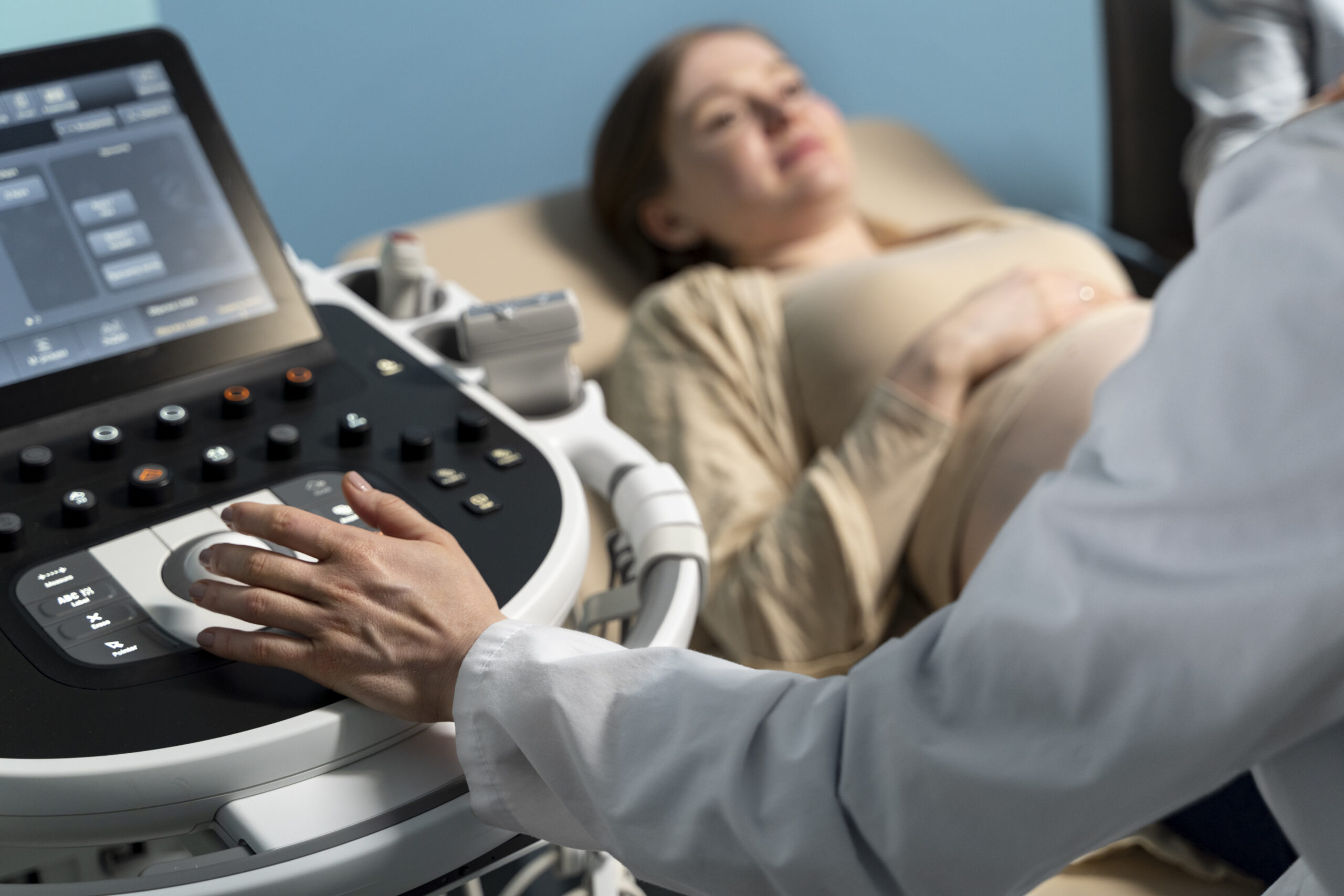
Pregnancy ultrasound or obstetric ultrasound

RDV IRM en ligne sur Doctolib
Prenez rendez-vous pour une IRM dans nos centres
Prise de rendez-vous IRM- Paris 75002
Prise de rendez-vous IRM – Paris 75009

Pregnancy ultrasound, also known as obstetrical ultrasound, is a common medical procedure used to monitor the development of the baby during pregnancy. It's a medical imaging method that doesn't require intervention and allows healthcare professionals and parents to see the baby inside the mother's womb. In this text, we'll talk about the importance of pregnancy ultrasound, its various uses and its important role in monitoring the health of both mother and baby.
Understanding obstetrical ultrasound
Obstetrical ultrasound is when special sound waves are used to take live pictures of the baby, the placenta and the mother-to-be's uterus. A specialist called an ultrasonographer, or a healthcare professional trained in the use of ultrasound equipment, usually performs this procedure. Ultrasound scans can be performed at various times during pregnancy to see how the baby is growing and developing.
Obstetrical ultrasound is a useful clinical examination for :
- établir la présence d’un embryon ou d’un fœtus vivant ;
- estimer l’âge de la grossesse ;
- diagnostiquer des anomalies congénitales du fœtus.
- assess fetal position
- assess the position of the placenta
- déterminer s’il y a des grossesses multiples ;
- déterminer la quantité de liquide amniotique autour du bébé.
- vérifier l’ouverture ou le raccourcissement du col de l’utérus.
- évaluer la croissance fœtale ;
- assess fetal well-being
Some doctors also use 3D ultrasound to visualize the fetus and determine whether it is developing normally.
The different phases of a pregnancy ultrasound
- Early ultrasound : habituellement, elle se fait au début de la grossesse pour confirmer qu’il y a bien une grossesse, pour voir combien de bébés il y a et pour estimer quand le bébé va naître. Cette première échographie aide aussi à repérer d’éventuels problèmes dès le début.
- Morphological ultrasound : elle se fait entre la 18ᵉ et la 22ᵉ semaine de grossesse. L’objectif est d’examiner la structure du bébé, comme son cœur, son cerveau, sa colonne vertébrale et ses membres, afin de détecter d’éventuels problèmes anatomiques.
- Growth ultrasound : celle-ci se fait au cours du dernier trimestre de la grossesse pour vérifier la croissance du bébé, la quantité de liquide autour de lui et la position du placenta. Cela permet de s’assurer que le bébé se développe bien et aide à prendre des décisions éclairées sur le suivi de la grossesse.
The importance of obstetrical ultrasound
- Early detection of problems Ultrasound during pregnancy helps to identify potential fetal problems early on, enabling parents to make informed decisions about how to manage the pregnancy.
- Monitoring the growth of the unborn baby : : en vérifiant régulièrement la croissance du fœtus, les professionnels de la santé peuvent repérer toute anomalie de croissance qui pourrait nécessiter une intervention médicale.
- Gender identification : même si beaucoup de parents voient cela comme un moment amusant pendant l’échographie, savoir le sexe du bébé peut aussi être important pour des raisons médicales, surtout en cas de maladies génétiques liées au sexe.
- Birth preparation : Ultrasound allows parents to see their unborn child, strengthening their emotional bond with him/her and helping them prepare for childbirth and parenthood.
Emerging technologies in obstetrical ultrasound
Over the years, technological advances have greatly improved the quality and precision of ultrasound images. 3D and 4D ultrasound techniques enable parents to see their baby more realistically, with greater facial and movement detail. These advances not only please parents, they can also provide useful information for medical follow-up.
Challenges and ethical considerations
Although a pregnancy obstetrical ultrasound is a common procedure, it is not without its challenges and controversies. Some healthcare professionals and ethical groups are raising concerns about the use of ultrasound for non-medical purposes, such as sex determination for prenatal selection. It is important to maintain a balance between the medical benefits of ultrasound and the need to respect parents' ethical and cultural choices.
Obstetrical ultrasound examination
À quoi ressemble l’équipement d’échographie obstétricale ?
The equipment used for this procedure typically includes a computer console, a video monitor and a transducer, which is a small hand-held device resembling a microphone. Different types of transducer can be used in the same examination, each with specific capabilities tailored to the needs of the assessment.
The transducer emits high-frequency sound waves, inaudible to the human ear, into the body, and listens to the returning echoes. This principle is similar to the sonar used by boats and submarines. During the examination, a technologist applies a small amount of water-based gel to the patient's skin over the area to be examined. This gel enables sound waves to travel efficiently between the transducer and the skin, eliminating any air pockets that might block the passage of the waves. Ultrasound images are immediately visible on the video monitor, created by a computer which analyzes the intensity, frequency and return time of the ultrasound signals, taking into account the different body structures the waves pass through.
How obstetrical ultrasound works
The procedure works on similar principles to those used by bats or fishermen, who use echoes to detect objects in their environment. When sound waves encounter an object, they bounce back to the transducer. By measuring these echoes, it is possible to determine the distance, size, shape and consistency of internal structures, whether solid or liquid-filled. Doctors use ultrasound to detect abnormalities in the appearance of organs, tissues and blood vessels, as well as to identify abnormal masses, such as tumors.
During an ultrasound examination, the transducer sends out sound waves while recording the returning echoes. When the transducer is pressed against the skin, it emits sound pulses, and echoes generated by internal organs, fluids and tissues are picked up by a sensitive receiver in the transducer. A computer instantly processes this data, displaying images in real time on the monitor. The technologist can capture still images or record short video sequences of moving images, enabling visualization of fetal development and heartbeat.
A particularly interesting aspect of obstetrical ultrasound is the use of Doppler technology. This special application of ultrasound measures blood flow through the fetal heart, blood vessels and umbilical cord. Doppler works on the principle that the movement of red blood cells alters the frequency of reflected sound waves, creating a distinct sound effect. Patients often describe this sound as a breath noise, enabling doctors to monitor blood circulation and assess fetal well-being.
How does the procedure work?
For most ultrasound examinations, the patient lies on her back on an examination table, which can be tilted to improve image quality. The radiologist or sonographer positions the patient and applies gel to the area to be examined. The sonographer is then gently moved over the skin to capture the necessary images. Although some pressure may be felt, the examination is generally painless. Once imaging is complete, the technologist cleans the gel from the skin, which evaporates quickly without staining clothing.
In some cases, a transvaginal examination may be required to obtain more detailed images of the uterus and ovaries, particularly in early pregnancy. This procedure is similar to a conventional gynecological examination, with the transducer inserted into the vagina after the patient has emptied her bladder. With this approach, the doctor can obtain images from different angles to better visualize pelvic structures.
Obstetrical ultrasound is therefore an essential tool for monitoring pregnancies, providing crucial information on the health of both fetus and mother. Thanks to technological advances, this examination has evolved to become a safe and effective method for closely monitoring fetal development and intervening rapidly when necessary.
Pourquoi choisir IMPC pour votre échographie de grossesse (échographie obstétricale) ?
-
10 centres d’imagerie modernes à Paris, facilement accessibles.
-
Équipements d’échographie de dernière génération, offrant une excellente qualité d’image pour le suivi de la grossesse.
-
Médecins radiologues et échographistes spécialisés en imagerie obstétricale, attentifs au bon déroulement de l’examen.
-
Prise de rendez-vous rapide, sur Doctolib ou par téléphone.
-
Compte rendu clair et immédiat, remis en mains propres ou disponible rapidement en ligne.
