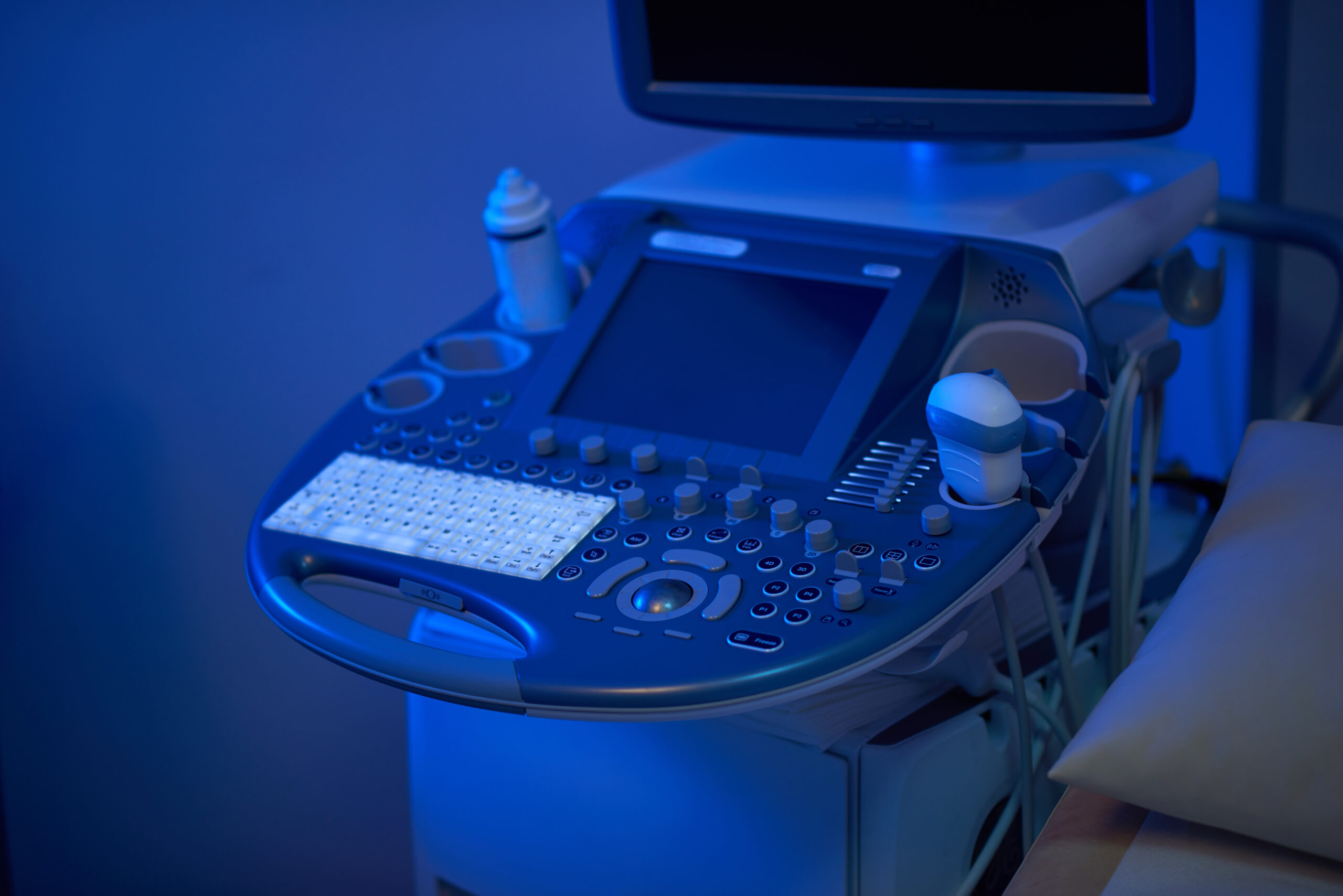
Hip ultrasound

RDV échographie en ligne sur Doctolib
Prenez rendez-vous pour un échographie dans nos centres
Prise de rendez-vous échographie – Paris 75009
Prise de rendez-vous échographie – Paris 75015
Prise de rendez-vous échographie – Paris 75009 Drouot

When it comes to diagnosing hip problems, ultrasound is a valuable tool. In this article, we'll explore hip ultrasound in depth, from its meaning and procedure to its benefits and limitations. If you are considering or need a hip ultrasound, read on for all the information you need.
What is a hip ultrasound?
A hip ultrasound is a medical imaging procedure that uses sound waves to create real-time images of the hip region. These images, called ultrasounds, enable healthcare professionals to visualize the internal structures of the hip, such as bones, joints, tendons and muscles. It's a non-invasive, painless tool that can help diagnose a variety of hip conditions.
Why might I need a hip ultrasound?
There are several reasons why a doctor may recommend a hip ultrasound. The main indications include:
- Hip pain : If you suffer from hip pain, an ultrasound can help determine the underlying cause, whether it's injury, inflammation or another condition.
- Joint problems : Ultrasound can detect joint abnormalities, such as bursitis or tendonitis, that affect the hip.
- Injury tracking : Ultrasound: Athletes and people who have suffered a hip injury may benefit from an ultrasound scan to assess the extent of damage.
- Muscle abnormalities, such as tears and soft-tissue masses.
- Foreign bodies, bleeding, infections or other types of fluid collection.
- Benign and malignant soft tissue tumors.
- Early changes in arthritis.
- Growth monitoring in infants Hip ultrasound in infants is used to detect and monitor developmental abnormalities, such as hip dysplasia. Developmental dysplasia of the hip (DDH), which in infants can vary from a shallow cup (bony acetabular dysplasia) to complete dislocation with the femoral head fully protruding from the acetabulum. Hip ultrasound can be performed in infants with DDH up to around six months of age.
Preparation before hip ultrasound
Before undergoing a hip ultrasound, there are a few preparation steps to follow. You may need to :
- Wear appropriate clothing : Wear comfortable, easy-to-remove clothing, as you may need to change your outfit for the examination.
- Remove jewelry and metal objects : You'll need to remove any metal objects that might interfere with the ultrasound sound waves.
- Follow the doctor's instructions : Your doctor will give you specific instructions on how to prepare for your particular situation.
How is a hip ultrasound performed?
Hip ultrasound is a relatively simple procedure that generally follows these steps:
Patient positioning
You will lie on an examination table, usually on your back, with the hip to be examined exposed.
Use of ultrasound gel
The technician will apply an ultrasound gel to the skin of the hip. This gel helps transmit sound waves and improves image quality.
The role of the technician
An ultrasound technician, also known as a sonographer, will manipulate an ultrasound probe over the hip area to obtain detailed images.
Pourquoi choisir IMPC pour votre échographie de la hanche ?
-
Un réseau de 10 centres d’imagerie médicale à Paris.
-
Des radiologues spécialisés en imagerie ostéo-articulaire et pédiatrique.
-
Des échographes modernes pour des images fiables et rapides.
-
Prise de rendez-vous facile sur la page contact ↗.
-
Une prise en charge personnalisée et rassurante.
Interpretation of results
Ultrasound images of the hip are immediately visible and interpreted by a radiologist or physician. They will look for abnormalities and diagnose common hip problems.
Anomaly detection
Ultrasound can detect various abnormalities, such as lesions, cysts, joint effusions or muscle problems.
Diagnosis of common hip problems
Hip ultrasounds are commonly used to diagnose conditions such as arthritis, bursitis and tendonitis.
Advantages and limitations of hip ultrasound
Hip ultrasound is a medical imaging technique commonly used to examine the hip region. It has both significant advantages and limitations to consider.
Préparation à l’examen
Pour optimiser la qualité des images, certaines consignes peuvent être données :
- Être à jeun pendant 4 à 6 heures dans certains cas.
- Ne pas vider complètement la vessie juste avant l’examen.
- Réaliser un lavement évacuateur 1 à 3 heures avant l’examen, notamment en cas de suspicion d’endométriose ou de pathologie rectale, afin d’éviter les artefacts d’image.
Injection de produit de contraste
Dans certains cas, une injection intraveineuse de gadolinium est réalisée. Elle permet de mieux visualiser certaines lésions ou tumeurs.
Les réactions allergiques sont extrêmement rares (<0,1 % des cas, HAS 2021).
Sécurité et contre-indications
L’IRM pelvienne est un examen sûr :
- aucun rayonnement ionisant,
- effets secondaires très rares,
- compatible avec la grossesse après le 1er trimestre (aucun effet nocif connu).
Contre-indications absolues
- Pacemaker non compatible.
- Implants auditifs ou certains dispositifs métalliques.
Tableau comparatif : échographie de la hanche vs IRM de la hanche
| Critère | Échographie de la hanche | IRM de la hanche |
|---|---|---|
| Précision diagnostique | Bonne pour tendons, muscles, bursites | Très élevée pour cartilages, labrum, os |
| Rayons X | Aucun | Aucun |
| Duration | 15–20 minutes | 20–40 minutes |
| Indications principales | Dysplasie, tendinites, bursites, épanchements | Arthrose, lésions cartilagineuses, fractures occultes |
FAQ : Les questions les plus fréquentes sur l’échographie de la hanche
Faut-il être à jeun pour une échographie de la hanche ?
Non, aucune préparation particulière n’est nécessaire.
L’échographie de la hanche est-elle douloureuse ?
Non, l’examen est totalement indolore, seule une légère pression de la sonde peut être ressentie.
À quel âge fait-on une échographie de la hanche chez le bébé ?
Elle est généralement réalisée entre 1 et 3 mois pour dépister la dysplasie.
Quand reçoit-on les résultats ?
Le radiologue commente les images immédiatement et remet un compte rendu écrit.
Sources scientifiques
-
Haute Autorité de Santé (HAS) – Imagerie musculo-squelettique et pédiatrique (2021).
-
Société Française de Radiologie – Recommandations en échographie de la hanche.
-
Inserm – Dépistage précoce des dysplasies de hanche chez le nourrisson (2022).
Dernière mise à jour : le 30 août 2025
Dr Anne Elodie Millischer
