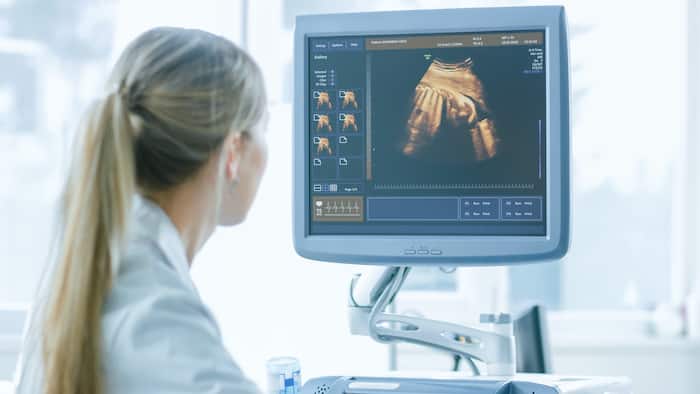
An ultrasound scan
Ultrasound in Paris
Ultrasound in Paris
What is an ultrasound scan?
Ultrasound is a painless, non-irradiating examination that uses the properties of ultrasound, without any consequences for the patient's health. The technology is similar to that used by sonar and radar, which help the military detect aircraft and ships. An ultrasound allows your doctor to see problems with organs, vessels and tissues without needing to make an incision.
Unlike other imaging techniques, ultrasound uses no radiation. For this reason, it is the preferred method for visualizing a developing fetus during pregnancy.

What is an abdomino-pelvic ultrasound?
Aultrasound abdomino-pelvic ultrasound enables visualization of the abdominal and pelvic organs. This examination is performed through the abdominal wall or by endocavity.
Why do I need an abdomino-pelvic ultrasound?
- Diagnosing a disease : tumor detection (e.g. ovarian cyst), infection (salpingitis, cholecystitis, pyelonephritis for example), a calculation (vesicular lithiasiskidney, for example)... ;
- Keeping pace of a pathology (cancer surveillance, treatment for a endometriosis…) ;
- Tracking growth and study the proper development of fetal organs during the pregnancy ;
- Guiding a surgeon through certain operationsetc.
When should I have an abdomino-pelvic ultrasound?
Pelvic pain, i.e. pain felt in the lower abdomen, which generally involves the uterus, bladder and rectum, and abdominal pain, which can take the form of cramping, burning or throbbing, can in some cases reveal abnormalities that require follow-up, treatment or intervention.
Any pelvic or abdominal pain persisting for more than 6 weeks should be reported to a radiologist.
What is an abdominal ultrasound ?
It's the ideal examination for exploring solid organs: the liver, gallbladder and bile ducts, kidneys and spleen.
Why do I need an abdominal ultrasound?
L'ultrasound abdominal provides a wealth of information, much richer than standard radiographyon the abdominal organs.
The main organs explored by abdominal ultrasound and the abnormalities are :
Gallbladder and bile ducts: stones, dilatation, tumors, abscesses, inflammation of the wall ;
Liver: size and homogeneity (cirrhosis, steatosis), cysts, abscesses, tumors, metastases of non-hepatic cancers;
Spleen: size, swelling, suspected rupture;
Pancreas: cyst, pancreatitis due to gallstones, cancer;
Kidneys: stones, dilated pelvis or ureters, tumours, atrophy, malposition, cysts, malformations;
Aorta and inferior vena cava;
The muscular wall of the abdomen.
Ultrasound may also show abnormal lymph nodes, ascites, blood or suspicious masses in the abdominal cavity.
When should I have an abdominal ultrasound?
If abdominal pain persists beyond 6 weeks, an ultrasound scan may be prescribed.
Do you have to fast before theabdominal ultrasound ?
To optimize the examination, you must be fasting and not have had a drink or smoked for at least 3 hours.
Ultrasound RDV
Make an appointment for an ultrasound scan at one of our centers:
MRI Bachaumont 75002
What is an obstetrical ultrasound?
Aultrasound Obstetrical ultrasound is a medical radiological examination that visualizes images of the baby (embryo or fetus) in a pregnant woman, as well as the mother's uterus and ovaries. It uses no ionizing radiation, has no known harmful effects and is the preferred method for monitoring pregnant women and their unborn babies.
That said, the use of obstetrical ultrasound is unquestionably one of the most essential examinations during pregnancy.
Why do I need an obstetrical ultrasound?
Aobstetrical ultrasound is a useful clinical examination for :
- Establishing the presence of a living embryo/foetus
- Estimating pregnancy age
- Diagnosing congenital anomalies of the fetus
- Assessing fetal position
- Assessing the position of the placenta
- Determining multiple pregnancies
- Determining the amount of amniotic fluid around the baby
- Check for cervical opening or shortening
- Assessing fetal growth
- Assessing fetal well-being
What's more, its usefulness is enhanced by the fact that this ultrasound assiduously accompanies the woman's entire pregnancy, paying particular attention to the baby's development right up to birth.
When should I have an obstetrical ultrasound?
In fact, three obstetrical ultrasounds are required during pregnancy.
- A dating ultrasound: this takes place at the very beginning. This is used to mark the date and make an appointment for ultrasound to be performed between 11 weeks and 1 day and 13 weeks and 6 days of amenorrhea.
- La 2e Ultrasound is a much more in-depth study of fetal morphology and adnexa (Doppler, placenta).
- La 3e The ultrasound will determine the baby's development, fetal weight, fetal position and placental position. The heart and brain will be more visible than on the 2e quarter.
How does an ultrasound scan work?
You'll be lying down in a darkened room to make it easier to read the images on the video screen, which you can follow on a second screen dedicated to the patient.
A gel will be applied to the skin to enable ultrasound transmission.
The examination provides dynamic moving images, controlled on a screen.
What is it all about?
Ultrasound uses ultrasound emitted by a probe and transmitted through the tissues, which reflect them, to form an image of the region examined. It can be coupled with a kind of radar to study vessels (Doppler).
Pelvic ultrasound
During thepelvic ultrasoundIf this is the case, do not urinate for 3 hours before the examination, or if you have urinated, drink 1 liter of water 1 hour beforehand.
To be in immediate contact with the area under examination and improve image quality, you may be offered the option of placing a probe covered with sterile protection in the rectum or vagina.
Very rarely, the introduction of the probe may cause transient, non-serious discomfort. Not all anatomical structures can be visualized by ultrasound. The liver, spleen, kidneys, bladder, uterus and ovaries are particularly well studied.
The study of the kidneys and bladder as well as gynecological organs often requires a full bladder examination.
Muscles and tendons are also well studied, but osteoarticular ultrasound requires no special preparation of the patient.
Is it necessary to inject a product?
NO, the test is performed without injection (prick) and is painless.
Abdominal ultrasound
During theabdominal ultrasoundYou must fast for 3 hours before your appointment, but take your usual medication.
Exploration will often require you to hold your breath for a few seconds.
Your results
An initial commentary may be given to you immediately after the examination, but this is only a first approach, as the images must then be the subject of a written report, which will be made available as soon as possible. It's normal to have questions about the examination you're about to undergo. We hope we've answered them. Please do not hesitate to contact us again if you require any further information.
